SOME ANATOMY TO BEGIN: There are three surfaces to a tendon
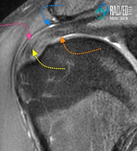
- Bursal ( Pink arrow)
- Intrasubstance ( Yellow Arrow)
- Articular ( Orange arrow)
The Blue arrow demonstrates the subacromial/ subdeltoid bursa
WHAT IS A FULL THICKNESS TEAR:
- We are assessing the tendon from the superior ( bursal) to its inferior ( articular) surface.
- A full thickness tear is a tear that involves the entire tendon in the superior to inferior direction.
- Another way to put it is that it extends all the way between the Articular and Bursal Surfaces.
Lets break it down to the individual words
FULL THICKNESS:
- We are assessing the tendon from its superior ( bursal) to inferior ( articular) surface.
- The tear extends from Bursal to Articular surface of tendon.
INCOMPLETE OR LOCALISED:
- We are now assessing the tendon in the Anterior to Posterior direction.
- Incomplete means, in the AP direction only a part of the tendon is torn.
COMPLETE:
- Complete means, in the AP direction the entire tendon is torn.
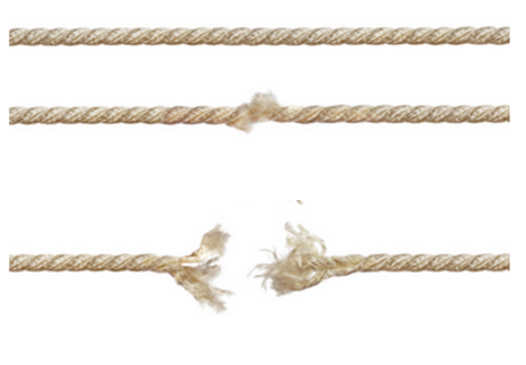
If you look at the image below of the rope and imagine its the Supraspinatus Tendon
- The first image the SST is intact and not torn.
- The second image would be a Full Thickness Incomplete/ Localised tear ( One part of the tendon has a full thickness tear whilst the rest of the tendon is still intact).
- The tird image would be a Full Thickness Complete Tear. ( The tendon in its entirity has a full thicknes stear.)
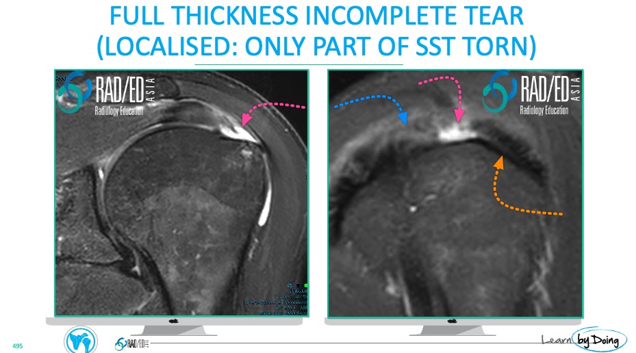
WHAT TO LOOK FOR:
- FULL THICKNESS: Look for high signal ( Fluid type) extending from bursal to articular surface of tendon ( Pink arrow in image below).
- INCOMPLETE/ LOCALISED: Involves only a portion of the tendon ( Pink arrow). The remainder of the SST ( Blue arrow) is intact. Orange arrow = IST
WHAT TO LOOK FOR:
- FULL THICKNESS: Look for high signal ( Fluid type) extending from bursal to articular surface of tendon( Blue arrows in image below).
- COMPLETE: Involves the entire SS tendon. No component of the SST is continuous and intact. The IST ( Yellow arrow) is intact.
- MTJ RETRACTION: With a complete, full thickness tear the musculo tendinous junction is retracted ( Pink Arrow) because the tendon is no longer attached to the greater tuberosity.

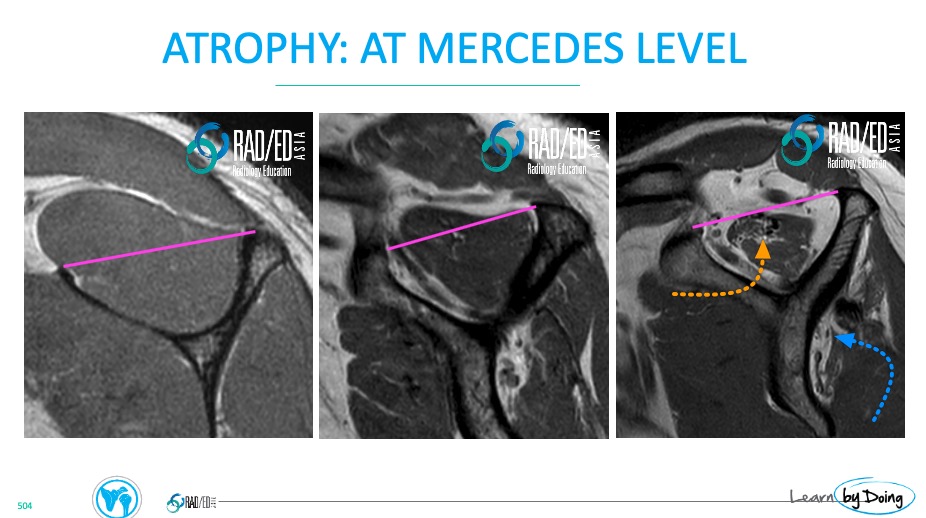
WHAT TO LOOK FOR:
4. ATROPHY +/- FATTY INFILTRATION OF THE MUSCLE.
- Atrophy and Fatty infiltration are important to report.
- The worse the atrophy and fatty infiltration, the less chance of a good functional outcome if surgery is performed.
- ATROPHY: Assess on Sagittal NON Fat Sat sequence, at level of the Mercedes Sign ( see below). There is no measurement
- Normal SS muscle should be above the pink line.
- Atrophy if muscle below level of pink line.
- Describe as mild, moderate or severe.
- FATTY INFILTRATION: Assess on Sagittal NON Fat Sat sequence
- Normal muscle belly has minimal fat ( high signal T1 or PD).
- Look for fat in muscle ( Orange arrow in SS and Blue arrow in IS).
- Varying amounts of fatty infiltration. Describe as mild, moderate, severe.