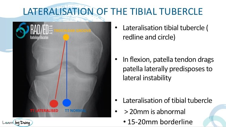
In Patellar Dislocation, Lateralisation of the position of the tibial tubercle is one of the predisposing factors. We look at what the normal position is, how to measure lateralisation and what it looks like.
| The Tibial Tubercle: Normal and Lateralised |
|
NORMAL:
LATERALISED:
The measurements above are based on CT evaluation where the knee is in full extension. However in MRI with a knee coil the knee is about 15 degrees of flexion which results in some medialisation of the tibial tubercle. In MRI Normal is considered <15mm. |
| How to Measure it |
|
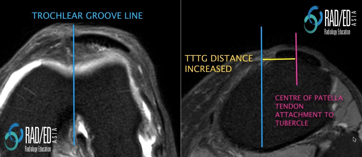
The transverse distance between the trochlear groove and the tibial tuberosity is called the TT-TG distance ( Tibial Tuberosity Trochlear Groove distance).
HOW DO YOU MEASURE IT ON A SCAN
|

| What Does Lateralisation of the tubercle look like? |
|
With lateralisation of the tibial tubercle, there is increased transverse distance between the centre of the trochlear groove and the central point of the patella tendon at its attachment to the tubercle.
|
| How do CT Vs MRI Measurements Compare |
|
The TT-TG distance can vary between CT and MRI and also within MRIs depending on the position of the knee.
(CT and MRI Measurements of Tibial Tubercle–Trochlear Groove Distances Are Not Equivalent in Patients With Patellar Instability American Journal of Sports Medicine Vol 41, Issue 8, 2013 ) |