There are a few intra articular normal variants that we will commonly see on MRI of the elbow. This post reviews the four most common variants that could be confused with pathology.
1. Trochlear Ridge
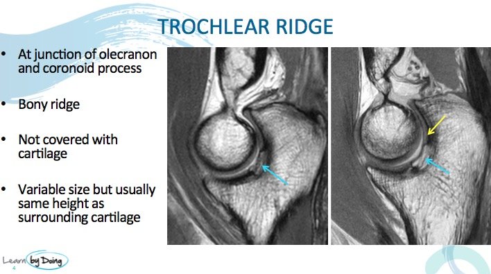 Image Above: Blue Arrow Trochlear ridge. Yellow arrow normal cartilage. Trochlear ridge not covered with cartilage but is approximately the same height as adjacent cartilage.
2. Pseudo Defect of the Trochlear
 Image above: Pseudo defect of the trochlea ( blue arrow). Localised defect in trochlear groove not covered by cartilage or bone.
3. Pseudo Defect of the Capitellum
 Image Above: Pseudo defect of the capitellum ( blue arrows). Flattening of the capitellum but covered by cartilage.
 Image above: Defect caused by OCD is more anterior than a pseudo defect.
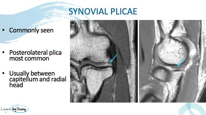
4. Synovial Plicae
UPCOMING MINI FELLOWSHIPS & WORKSHOPS


