Elbow MRI Brachialis tendon tendinosis and tears.
The Brachialis tendon is less commonly injured than the biceps. It inserts onto the anterior ulnar on the ulnar tuberosity and to a lesser extent on the coronoid process but the tendon is very short compared to the biceps tendon. Most commonly we see tendinosis or a strain/ partial tear at the musical tendinous junction. Complete ruptures are uncommon.
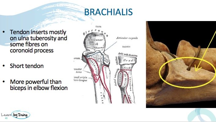
Image Above: Location of insertion of brachialis tendon. ( Image credit First Image Bartleby.com: Gray’s Anatomy, Plate 213, 2nd Image Source unknown please inform us if this is yours and we will acknowledge)
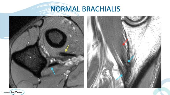
Image Above: Normal Brachialis tendon ( blue arrow). Should be black on all sequences like all tendons. Second image sagittal scan, broad tendon attachment ( blue arrow) with a very short tendon length. Brachialis muscle ( red arrow). Biceps tendon ( yellow arrow).
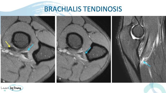
Image above: Brachialis tendinosis standard appearance of tendinosis in any tendon with increased PDFS signal in tendon and peritendinous oedema ( blue arrow). Mild tendinosis biceps tendon ( yellow arrow).

Image above: Brachialis tendon ( yellow arrow) barely seen due to tendinosis and delamination. Compare signal with normal biceps tendon ( blue arrow).

Image Above: Brachialis musculotendinous junction ( MTJ) strain and partial tearing. Oedema in muscle at MTJ ( blue arrow). Brachialis tendon ( yellow arrow) appears normal.
UPCOMING MINI FELLOWSHIPS & WORKSHOPS ( Kuala Lumpur is Full )


