ROTATOR CUFF ANATOMY

Before we look at the pathology lets start with anatomy. We will concentrate on SST and IST and will do Subscapularis in a different post.

Because there is no plane between the SST and IST tendons, one of the difficulties is in determining on MRI which tendon is involved when there is a tear at the insertion region. We can make this easier by

  1. Understanding the insertion sites on the greater tuberosity and by
  2. Understanding the different muscle shapes and following them.

ANATOMY OF THE GREATER TUBEROSITY

The SST and IST tendons both attach to the greater tuberosity which has 3 facets where three tendons attach

rct facet insertions

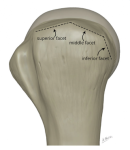
  1. Horizontal/ Superior Facet: SST Attaches ( Green Dashed Line in image below)
  2. Oblique/ Middle Facet IST Attaches ( ( Pink Dashed Line in image below)
  3. Vertical/ Inferior Facet Teres Minor Attaches ( Blue Dashed Line in image below)

USING MUSCLE SHAPE

The Supraspinatus and Infraspinatus have different shapes.

  1. On Coronal scans the SS is anterior and more ovoid in shape and the IS is posterior and flatter.
  2. On Sagittal Scans the SS lies more superiorly and the IS more posteriorly and inferiorly.

You can use the muscle bellies to work out which tendon is involved at the insertion by following the muscle down to its insertion.


WHAT IS IT: One important part of the anatomy is understanding that the SST has a very anterior insertion which we often dont look at properly and can miss tears at this region.

The anterior most part of the SST insertion is also called the Leading Edge.

WHERE TO LOOK FOR IT:

  1. On coronal scans find the biceps tendon ( pink arrow in image below).
  2. At this level the superior most margin of the groove is where the anterior most fibres of the SST insert ( Blue arrows in image below).
  3. To assess the SST, begin looking from the level of the biceps tendon.

Image Above: Blue arrows the anterior most fibres of SST. Pink arrows bBiceps tendon at bicipital groove.

Leave a Comment

error: Right Click not available.
Scroll to Top

This site is intended for Medical Professions only. Use of this site is governed by our Terms of Service and Privacy Statement which can be found by clicking on the links. Please accept before proceeding to the website.