
WHAT IS IT: Fraying is irregularity of the surface of the SST or IST. It is not a tear.
WHAT AND WHERE TO LOOK FOR:
- Fraying occurs on the BURSAL SURFACE of the SST or IST.
- Look for an irregular margin of the tendon.
- The best analogy is to think of fraying like scratches on the skin. The surface becomes irregular but there is no tear to stitch.
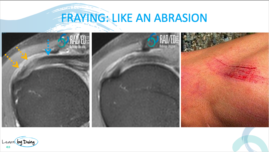
Image Above: Blue arrows normal sharp margin of the bursal Orange arrows irregular margin of the tendon in keeping with fraying.
WHAT IS IT: Tendonosis is degeneration of the tendon. It is not a tear.
WHAT AND WHERE TO LOOK FOR:
- A normal tendon is black. In tendonosis there will be an increase in tendon signal on PD or PDFS sequences.
- The signal increase is intermediate, not really bright like fluid.
- The tendon may be increased in size.
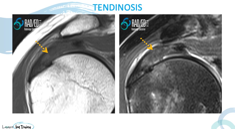
Image Above: Orange arrows increased signal in tendon but signal is not fluid signal. Tendon is also slightly thickened.
HOW DO YOU DIFFERENTIATE TENDONOSIS FROM TEARS:
We will go into the appearance of tears in posts to come but the main difference is signal on PDFS or T2FS sequences.
- Tendonosis is intermediate signal ( orange arrow in image below)
- Tear are much brighter and are fluid signal ( Blue arrow in image below).
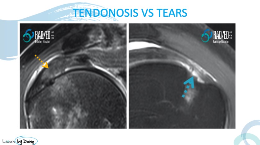
Image Above: Tendonosis ( Orange Arrow) vs Tear ( Blue arrow). A tear is much higher in signal and is of fluid signal.
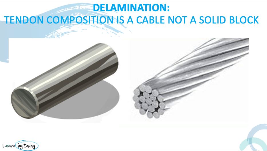
WHAT IS IT: A tendon is like a wire cable, composed of multiple fibres rather than a solid block like a steel rod.
Delamination is a longitudinal separation of tendon fibres. Its as if the fibres peel off each other and separate with the area of separation in between mucoid material. It is not a tear.
WHAT AND WHERE TO LOOK FOR:
- Delamination is a severe form of degeneration.
- In delamination there will be longitudinal increase in tendon signal ( paralleling tendon fibres) on PD or PDFS sequences.
- The signal increase is intermediate, not really bright like fluid.
- The tendon may be increased in size from underlying tendonosis.
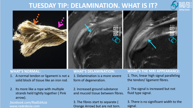
Image Above: Orange arrows increased signal in tendon but signal is not fluid signal. Tendon is also slightly thickened.
HOW DO YOU DIFFERENTIATE DELAMINATION FROM TEARS:
This can be a little subjective but what we are looking for is
- Increased signal ( not fluid) running parallel to the tendon fibres.
- There is no sense that the ares of increased signal have an significant volume to them like a tear.
- We will go more into this at the workshop.